
✨ This article was AI edited. Editorial responsibility: Hysteroscopy.info.
Produced with AI. This article was artificially generated. Editorial responsibility: Hysteroscopy.info.
A diagnostic hysteroscopy typically takes between 5 to 15 minutes, whereas an operative hysteroscopy (such as removing endometrial polyps, submucosal fibroids, or uterine adhesions) requires 15 to 45 minutes depending on surgical complexity. Including preoperative check-in, cervical preparation, local or general anesthesia administration, and post-anesthesia recovery observation, patients should anticipate a total clinic or surgical facility stay of approximately 1.5 to 3 hours.
When preparing for an endoscopic uterine examination or therapeutic intervention, one of the most pressing questions patients ask their gynecologist is: how long does a hysteroscopy procedure actually take? While the time spent actively visualizing or treating the uterine cavity is surprisingly brief, understanding the complete clinical timeline—from arrival and sedation to postoperative discharge and home recovery—is essential for minimizing anxiety and planning your schedule effectively.
Whether you are scheduled for an in-office diagnostic evaluation for abnormal uterine bleeding or an outpatient operative resection of structural pathology, this clinical guide breaks down the precise duration of every procedural stage, the medical variables that influence surgical time, and what milestones to anticipate during recovery.
At a Glance: Procedure Duration by Clinical Category
The total duration of a hysteroscopic procedure depends primarily on whether the intervention is purely observational (diagnostic) or interventional (operative). The table below outlines standard clinical durations across typical gynecological presentations:
| Procedure Variant | Typical Anesthesia | Active Surgical Time | Total Facility Stay | Return to Desk Work |
|---|---|---|---|---|
| Diagnostic Hysteroscopy (Office) | None or Local Paracervical Block | 5 – 10 minutes | 45 – 60 minutes | Same day or next morning |
| Diagnostic + Directed Endometrial Biopsy | Local Block or Oral Sedation | 10 – 15 minutes | 60 – 90 minutes | Next day |
| Operative Polypectomy (Endometrial Polyp) | Conscious Sedation or General Anesthesia | 15 – 25 minutes | 2 – 3 hours | 1 – 2 days |
| Operative Myomectomy (Submucosal Fibroid) | Monitored Anesthesia Care (MAC) or General | 25 – 45 minutes | 3 – 4 hours | 2 – 4 days |
| Lysis of Adhesions (Asherman’s Syndrome) | General Anesthesia | 30 – 60 minutes | 3 – 5 hours | 3 – 5 days |
For a detailed medical walkthrough of instrumentation and pre-surgical checks, explore the complete hysteroscopy process from initial consultation to discharge.
Step-by-Step Clinical Timeline: What Happens During Each Phase
Understanding the actual breakdown of time inside the medical facility demystifies the procedure. While the camera is inside the uterus for only a fraction of the appointment, several sequential phases ensure clinical safety, sterile technique, and patient comfort.
Phase 1: Pre-Procedure Intake and Cervical Preparation (30 to 45 Minutes)
Upon arrival at the ambulatory surgical center or specialized gynecology suite, intake procedures commence. A specialized nurse will record your baseline vitals (blood pressure, heart rate, oxygen saturation) and confirm pregnancy test results if premenopausal.
- Cervical Softening Evaluation: If you were prescribed oral or vaginal misoprostol 4 to 12 hours prior to the procedure to soften and prime the cervix, the clinician will evaluate cervical compliance.
- Intravenous Access & Sedation Setup: For operative procedures or patients electing conscious sedation, an IV line is established, and baseline prophylactic medications (such as IV non-steroidal anti-inflammatories or antiemetics) are administered.
- Pre-Operative Timeout: The surgeon, anesthesiologist, and nursing staff perform the formal surgical safety checklist confirming the patient identity, procedure indication, and allergy profile.
Phase 2: Patient Positioning, Disinfection & Anesthesia Induction (5 to 10 Minutes)
You will be escorted into the procedure room and positioned comfortably in dorsal lithotomy position with stirrups supporting your legs.
- Perineal Cleansing: The external genitalia, vaginal vault, and ectocervix are prepped with an antiseptic solution (such as povidone-iodine or chlorhexidine gluconate).
- Anesthesia Administration: For in-office diagnostic exams, a local paracervical block (injecting 1% or 2% lidocaine into the cervical stroma at the 4 and 8 o’clock positions) takes roughly 2 to 3 minutes to achieve deep local analgesia. If undergoing general anesthesia or deep IV propofol sedation, the anesthesiologist will administer medications, leading to restful sleep within 60 seconds.
Phase 3: Uterine Access and Cavity Distension (2 to 4 Minutes)
Before the uterine cavity can be inspected, it must be gently expanded. Under normal anatomical conditions, the anterior and posterior muscular walls of the uterus touch each other in a potential cavity.
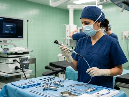
- Gentle Cervical Dilatation: Using graduated Hegar or Pratt cervical dilators, the cervical canal is dilated just enough to accommodate the hysteroscope sheath (typically 3 mm to 5.5 mm for diagnostic, or 7 mm to 9 mm for operative resectoscopes). In nulliparous patients or postmenopausal women with stenotic canals, this step requires delicate calibration.
- Fluid Distension: Sterile normal saline (0.9% sodium chloride) is instilled through the inflow channel of the sheath under controlled pressure (usually maintained between 70 to 100 mmHg). This distends the cavity, washes away micro-debris or mucus, and provides a crystal-clear panoramic view of the endometrium and bilateral tubal ostia.
To understand the precision equipment and medical safety protocols involved, review our comprehensive review on how is a hysteroscopy done step-by-step.
Phase 4: Direct Endoscopic Inspection and Operative Intervention (5 to 40 Minutes)
This is the core phase of the procedure where diagnostic answers are established or therapeutic pathology is excised.
- Diagnostic Inspection (5–10 mins): The gynecologist systematically scans the endocervical canal, the internal cervical os, the lower uterine segment, the fundus, the anterior and posterior endometrial surfaces, and the right and left fallopian tube openings (ostia). If micro-polyps or localized hyperplasia are identified, targeted punch biopsies are retrieved using miniature grasping forceps passed through the operating channel.
- Operative Resection (15–40 mins): If an endometrial polyp or submucosal myoma is present, a mechanical tissue removal device (such as a hysteroscopic morcellator) or a bipolar electrosurgical loop resectoscope is introduced. The surgeon methodically shaves and vaporizes the lesion flush with the myometrial junction while continuously aspirating tissue chips for histological evaluation.
Phase 5: Instrument Withdrawal and Immediate Post-Op Recovery (30 to 60 Minutes)
Once the pathology is removed and hemostasis is verified, the distension fluid is completely evacuated, and the hysteroscope is withdrawn.
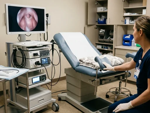
- Office Hysteroscopy Recovery: If performed under local block, you will rest in the exam chair for 10 to 15 minutes to ensure no vasovagal lightheadedness occurs before dressing and reviewing preliminary photographic findings with your doctor.
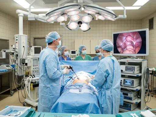
- Operating Room Post-Anesthesia Care Unit (PACU): If general anesthesia or IV sedation was used, you will be monitored in the recovery suite for 45 to 90 minutes while the anesthesia clears. Nurses continuously monitor blood pressure, assess post-procedural cramping, and provide a light beverage or snack.
Key Clinical Factors Influencing Procedure Duration
Every woman’s reproductive anatomy is unique, and several clinical nuances determine whether an operative procedure takes 15 minutes or extends toward 45 minutes:
1. Pathology Size, Number, and Vascularity
A solitary pedunculated endometrial polyp measuring 8 millimeters can be resected and retrieved in under 4 minutes. In contrast, a 3-centimeter broad-based submucosal fibroid (FIGO Type 1 or Type 2) with deep myometrial extension requires incremental mechanical shaving, careful vessel coagulation, and frequent monitoring of the fluid deficit to ensure complete patient safety.
2. Cervical Anatomy and Stenosis
Entering the cervical canal can take less than 30 seconds in a woman who has delivered vaginally. However, patients with a history of cervical excisional procedures (such as LEEP or cold knife cone biopsies), severe cervical scarring, or postmenopausal atrophy may have a tight, pinpoint external os. Gynecologists take extra time with sequential micro-dilators or acoustic ultrasound guidance to prevent creating a false passage or cervical laceration.
3. Choice of Anesthesia and Facility Setting
In-office ‘see-and-treat’ hysteroscopy using modern miniaturized 3.8 mm scopes eliminates the administrative and physiological delay of general anesthesia, allowing patients to complete the entire visit in less than 45 minutes. Hospital-based ambulatory surgery involves rigorous pre-operative documentation, regional or endotracheal anesthesia, and structured PACU recovery discharge criteria.
4. Fluid Management and Distension Medium Monitoring
During operative hysteroscopy, surgeons utilize automated fluid management systems to continuously calculate the difference between fluid instilled into the uterus and fluid recovered in the suction canisters. If the systemic fluid deficit reaches safety thresholds (typically 2,500 mL for isotonic normal saline or 1,000 mL for hypotonic non-electrolyte media), the procedure is paused or concluded to eliminate any risk of fluid overload or electrolyte imbalances.
To learn more about intraoperative patient monitoring and fluid safety, see our analysis of fluid management and distension media safety in hysteroscopy.
Post-Procedure Recovery Timeline: What to Expect at Home
Once discharged from the medical suite, recovery progresses rapidly. Because hysteroscopy is a true natural-orifice endoscopic procedure with zero external skin incisions, the physical healing curve is substantially faster than laparoscopy or open abdominal surgery.
Home Recovery Milestones:
- Hours 1 to 6 Post-Procedure: Mild to moderate menstrual-like uterine cramping. This is caused by the uterus contracting down as distension fluid empties and prostaglandins are released. Standard over-the-counter NSAIDs (such as 400–600 mg ibuprofen) or prescribed antispasmodics provide excellent relief.
- Days 1 to 3 Post-Procedure: Light vaginal spotting or water-pink serosanguinous discharge. Cramping typically resolves within 24 to 36 hours. Most women return to work, driving, and light desk duties within 24 to 48 hours.
- Days 4 to 10 Post-Procedure: Discharge gradually shifts from pale red to yellowish-brown before ceasing. Strenuous cardiovascular workouts, heavy lifting over 20 pounds, and abdominal straining can usually be resumed after 3 to 5 days.
For explicit clinical milestones regarding bathing, sexual intercourse, and physical exertion, refer to our detailed guide on how long to wait after hysteroscopy for physical activities.
Normal Discharge vs. Warning Signs
While light bleeding and intermittent spotting are entirely physiological after biopsy or polypectomy, patients should monitor their symptoms diligently. As detailed in our clinical protocol on bleeding after hysteroscopy: normal vs warning signs, contact your surgical team immediately if you experience:
- Heavy vaginal hemorrhage that saturates more than two maxi-pads per hour for two consecutive hours.
- Severe, escalating pelvic or abdominal pain that does not respond to oral analgesics.
- A persistent oral body temperature elevation exceeding 100.4°F (38.0°C) or chills.
- Malodorous or purulent vaginal discharge indicative of pelvic infection.
Frequently Asked Questions About Hysteroscopy Duration
How long will I be asleep if I have general anesthesia for hysteroscopy?
When general anesthesia or monitored anesthesia care (MAC) with IV propofol is administered, patients are typically asleep for 20 to 45 minutes in total. The anesthesiologist administers short-acting agents calibrated precisely to the estimated surgical duration. Awakening in the operating room occurs within 5 to 10 minutes following the conclusion of surgery, with full mental lucidity returning over the next 45 minutes in the recovery area.
Can I drive myself home after a hysteroscopy?
If you undergo an office diagnostic hysteroscopy without sedation or under local paracervical block only, you are medically permitted to drive yourself home once you feel fully comfortable. However, if any oral sedatives (e.g., diazepam), IV narcotics, or general anesthesia were utilized, hospital accreditation regulations strictly mandate that a designated adult companion escort you home, and driving is strictly prohibited for 24 hours.
Does hysteroscopy take longer if combined with a D&C (Dilation and Curettage)?
Combining a hysteroscopy with a dilation and curettage adds approximately 5 to 10 minutes to the total operating room time. During this combined procedure, the surgeon first inspects the cavity endoscopically, performs the curettage to collect endometrial samples or clear hyperplastic tissue, and frequently re-inserts the camera for a final visual audit to confirm that the uterine cavity is clean and hemostatic.
How soon will I receive biopsy results after the procedure?
While your surgeon can share immediate visual findings with you in the recovery room, any excised polyps, fibroid fragments, or endometrial curettings are submitted to surgical pathology for microscopic histological analysis. Formal pathology reports are typically completed and finalized within 5 to 7 business days.
Get our latest guides, news, and insights highlighted in your Google Search & AI Overviews.