
✨ This article was AI edited. Editorial responsibility: Hysteroscopy.info.
Produced with AI. This article was artificially generated. Editorial responsibility: Hysteroscopy.info.
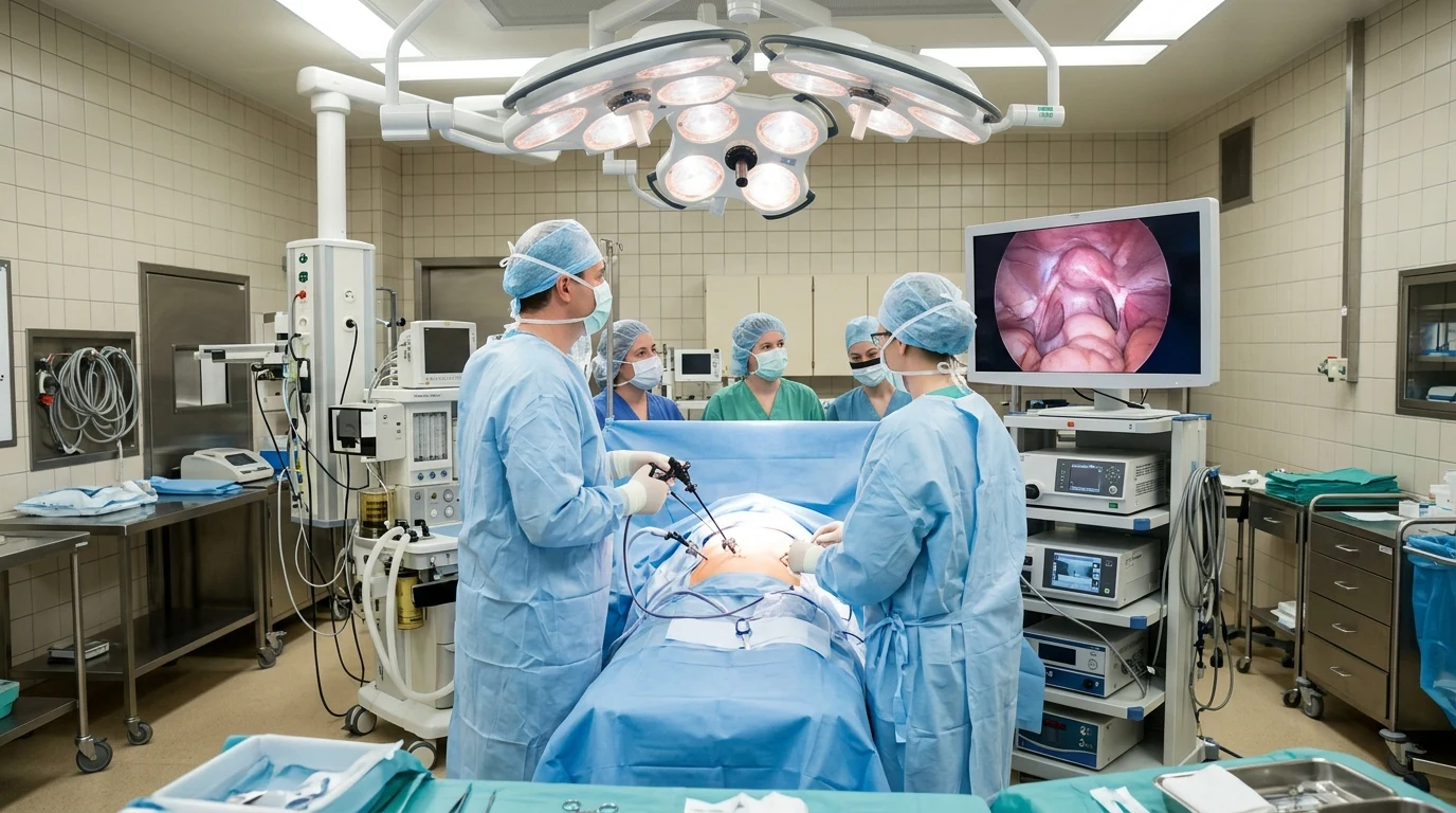
Hysteroscopic removal of fibroids (hysteroscopic myomectomy) is a transcervical, incision-free surgical procedure designed to resect submucosal uterine fibroids protruding into the endometrial cavity. Utilizing a resectoscope with a high-frequency bipolar wire loop or mechanical morcellation system, surgeons slice and extract fibroid tissue under continuous fluid distension, significantly reducing abnormal heavy uterine bleeding and enhancing reproductive capacity.
Submucosal uterine fibroids (leiomyomas) develop immediately beneath the endometrial lining and project into the uterine cavity. Despite representing only 5% to 10% of all uterine fibroids, submucosal fibroids are disproportionately responsible for severe menorrhagia (heavy menstrual bleeding), iron-deficiency anemia, dysmenorrhea, recurrent pregnancy loss, and embryo implantation failure. Hysteroscopic myomectomy provides a definitive, uterus-preserving surgical resolution without any skin incisions.
FIGO Classification of Submucosal Fibroids
Surgical feasibility and procedural planning for hysteroscopic fibroid removal depend directly on the International Federation of Gynecology and Obstetrics (FIGO) classification of submucosal leiomyomas:
| FIGO Subtype | Intramural Extension | Cavity Protrusion | Surgical Approach & Complexity |
|---|---|---|---|
| Type 0 | 0% (Pedunculated) | 100% within the cavity | Single-stage hysteroscopic resection; straightforward excision |
| Type 1 | < 50% intramural extension | ≥ 50% intracavitary protrusion | Standard hysteroscopic myomectomy; high success in one stage |
| Type 2 | ≥ 50% intramural extension | < 50% intracavitary protrusion | Advanced hysteroscopic surgery; may require staged two-step resection |
Surgical Modalities and Instrumentation
Modern hysteroscopic myomectomy employs two primary technological methodologies:
1. Classical Resectoscopy (Bipolar Wire Loop Electrosurgery)
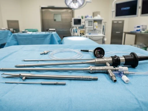
A rigid continuous-flow resectoscope equipped with a motorized semicircular bipolar wire loop is used to systematically shave the fibroid tissue from the dome down to its base in thin progressive chips (resembling a carpenter’s plane). The electrical current operates between two active poles on the probe tip within isotonic 0.9% normal saline, ensuring precise hemostatic cutting while eliminating the historical risks of hypotonic fluid-induced hyponatremia.
2. Hysteroscopic Tissue Removal Systems (Mechanical Morcellation)
Advanced mechanical tissue extraction systems (such as MyoSure or TruClear) utilize a rapidly rotating and reciprocating inner blade housed within a cannula. As the blade morcellates the fibroid tissue mechanically, integrated continuous vacuum suction instantly evacuates the tissue fragments out through the scope. This eliminates the need for repeated scope insertions and withdrawals, significantly reducing operating room time and mucosal trauma.
Distension Fluid Safety and Management
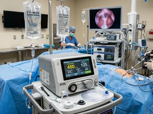
Intrauterine distension with normal saline is required to create an optical surgical field. During the shaving of myometrial tissue, open venous sinuses can absorb fluid into the systemic circulation. Intraoperative fluid management protocol dictates:
- Automated Fluid Balance Monitoring: Real-time electronic scale fluid measurement systems continuously track fluid inflow versus outflow to calculate the exact systemic absorption deficit.
- Maximum Allowable Deficit: In healthy premenopausal women undergoing bipolar resection with normal saline, the maximum fluid deficit is strictly capped at 2,500 mL. If the deficit reaches this limit, the procedure is immediately concluded to prevent pulmonary edema or fluid overload, and any residual intramural fibroid tissue is scheduled for a second-stage procedure.
Recovery Timeline and Post-Operative Expectations
Because hysteroscopic myomectomy is performed entirely through natural anatomical pathways (the cervix) without abdominal incisions, patient recovery is remarkably swift:
| Post-Operative Phase | Expected Symptoms | Clinical Management & Restrictions |
|---|---|---|
| Hours 0 – 6 | Mild lower abdominal cramping, grogginess from anesthesia, light reddish-brown spotting | Oral NSAIDs, rest in recovery area, discharge home with adult escort |
| Days 1 – 3 | Minimal pelvic discomfort, mild fatigue, pinkish or serosanguinous discharge | Return to light daily activities and remote work; avoid heavy lifting (>15 lbs) |
| Weeks 1 – 2 | Resolution of cramping; tapering light vaginal discharge as endometrial bed heals | Return to full physical exercise and work; complete pelvic rest (no intercourse, tampons) |
| Weeks 4 – 6 | First post-procedural menstrual period (often significantly lighter) | Post-operative clinical follow-up; clearance to attempt pregnancy or fertility treatments |
Clinical Efficacy for Bleeding and Fertility
Extensive clinical studies demonstrate outstanding patient outcomes following hysteroscopic fibroid removal:
- Menorrhagia Resolution: More than 85% to 92% of patients experience a dramatic reduction in menstrual blood loss, with restoration of normal hemoglobin levels and elimination of chronic anemia.
- Fertility Enhancement: Removal of cavity-distorting Type 0 and Type 1 submucosal fibroids increases clinical pregnancy and live birth rates by 40% to 50% in women experiencing unexplained infertility or recurrent IVF implantation failure.
- Uterine Integrity: Because the outer uterine myometrium and serosa remain untouched, women who undergo hysteroscopic myomectomy are generally candidates for normal vaginal deliveries in future pregnancies, unlike those who undergo open abdominal or laparoscopic transmyometrial myomectomies.
Frequently Asked Questions
Can uterine fibroids grow back after hysteroscopic removal?
The specific fibroid tissue resected during hysteroscopic myomectomy is permanently removed and cannot regrow. However, new fibroids can potentially develop in other areas of the myometrium over time. The overall recurrence rate requiring secondary intervention is approximately 10% to 15% over a 5-year period.
What type of anesthesia is used for hysteroscopic fibroid removal?
Most operative hysteroscopic myomectomies are performed under general anesthesia or monitored intravenous deep sedation (conscious sedation) with a paracervical block to ensure patient comfort and complete immobility during delicate intrauterine micro-resection.
How long after hysteroscopic myomectomy can I try to get pregnant?
Most gynecologists recommend waiting 1 to 2 complete normal menstrual cycles (approximately 6 to 8 weeks) to allow the endometrial cavity and myometrial resection bed to achieve complete epithelialization before attempting conception or initiating embryo transfer.
For more details on medical coding and pricing, read our CPT Code and Insurance Guide and our guide on Myomectomy vs. Hysteroscopy Options.
Get our latest guides, news, and insights highlighted in your Google Search & AI Overviews.