
✨ This article was AI edited. Editorial responsibility: Hysteroscopy.info.
Produced with AI. This article was artificially generated. Editorial responsibility: Hysteroscopy.info.
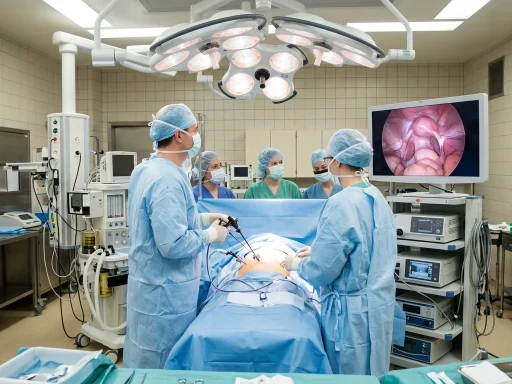
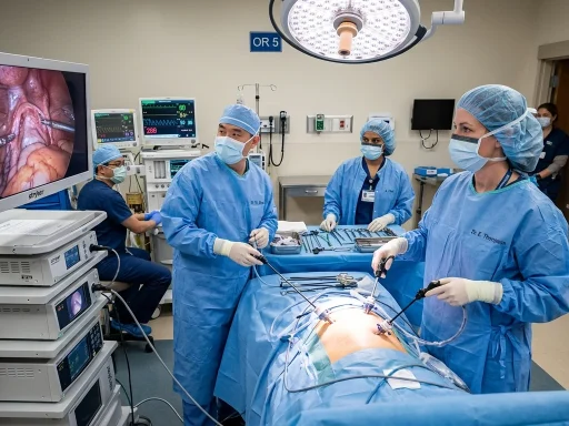
Hysteroscopic lysis of adhesions is a minimally invasive surgical procedure used to dissect and remove intrauterine scar tissue (synechiae), commonly associated with Asherman’s syndrome. Performed using micro-scissors or bipolar electrosurgery under direct endoscopic visualization, the procedure restores the normal anatomical shape and volume of the uterine cavity to alleviate hypomenorrhea, pelvic pain, and infertility.
Intrauterine adhesions (IUAs), also known as Asherman’s syndrome, develop when trauma to the basal endometrial layer triggers the formation of fibrous bands between the opposing uterine walls. Hysteroscopic adhesiolysis is the definitive gold-standard treatment, providing targeted anatomical restoration while preserving the surrounding healthy endometrium necessary for reproductive implantation and normal menstrual flow.
Etiology and Classification of Intrauterine Adhesions
The majority of intrauterine adhesions arise following postpartum or post-abortion curettage, operative hysteroscopy (such as myomectomy), or chronic intrauterine infections like pelvic inflammatory disease or genital tuberculosis. Intrauterine adhesions are clinically categorized by severity according to the American Society for Reproductive Medicine (ASRM) and European Society of Gynaecological Endoscopy (ESGE) staging systems:
| Classification Stage | Adhesion Characteristics | Uterine Cavity Occlusion | Prognostic Reproductive Impact |
|---|---|---|---|
| Mild (Stage I) | Thin, filmy, mucosal or avascular adhesions | Less than one-third (<33%) of cavity involved | Excellent spontaneous pregnancy prognosis (>80%) |
| Moderate (Stage II) | Thick, fibro-muscular bands; tubal ostia partially visible | One-third to two-thirds (33% – 66%) of cavity involved | Favorable with postoperative hormonal therapy (60% – 70%) |
| Severe (Stage III / IV) | Dense, fibrotic, confluent scar tissue; obliterated ostia | Greater than two-thirds (>66%) or complete cavity occlusion | Guarded; high recurrence risk; may require staged procedures |
Surgical Techniques for Hysteroscopic Adhesiolysis
The primary surgical objective of lysis of adhesions is the precise division of fibrotic bands from the central lower uterine segment upward toward the fundus and lateral cornua, avoiding false passages or uterine perforation. Three main surgical modalities are utilized:
1. Cold Scissor Dissection (Mechanical Micro-Scissors)
Cold scissors are widely regarded as the premier first-line tool for moderate to severe Asherman’s syndrome. Because no thermal or electrical energy is transmitted to the surrounding tissues, cold scissor adhesiolysis eliminates the risk of thermal necrosis to the remaining healthy basal endometrium, maximizing endometrial regenerative potential.
2. Bipolar Electrosurgery (Miniature Needle or Twizzle Electrodes)
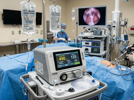
When dense, calcified, or fibrous myometrial adhesions resist mechanical division, miniature bipolar electrosurgical probes operating in isotonic normal saline provide controlled cutting. Energy settings are kept minimal to prevent collateral thermal spread into the underlying myometrium.
3. Concurrent Laparoscopic or Transabdominal Ultrasound Guidance
In cases of severe cavity obliteration where anatomical landmarks and tubal ostia are completely obscured, real-time transabdominal ultrasound or concurrent laparoscopy is employed. This provides direct visualization of the uterine wall thickness and prevents iatrogenic fundal perforation.
Post-Operative Prevention of Adhesion Recurrence
Intrauterine adhesions have a high propensity for re-formation during the initial 2 to 6 weeks of tissue healing, with recurrence rates exceeding 30% to 40% in severe cases. Multimodal anti-adhesion strategies are universally implemented immediately following surgery:
- Physical Barriers (Intrauterine Balloon / Stent): A pediatric Foley catheter or specialized triangular silicone intrauterine stent (e.g., Cook balloon) is placed in the cavity for 5 to 7 days to keep the anterior and posterior walls physically separated while the endometrial lining proliferates.
- Cross-Linked Hyaluronic Acid Gel: Anti-adhesive barrier gels (such as auto-crosslinked hyaluronic acid) are instilled into the cavity at the conclusion of the procedure to form a temporary protective macromolecular shield.
- High-Dose Estrogen & Progestin Therapy: Oral micronized estradiol (2mg to 4mg twice daily) is administered for 21 to 30 days to stimulate rapid endometrial proliferation and vascularization over the denuded myometrium, followed by a 10-day course of medroxyprogesterone acetate to induce withdrawal shedding.
- Second-Look Office Hysteroscopy: A follow-up diagnostic hysteroscopy is routinely scheduled 4 to 8 weeks post-surgery to inspect the healing cavity, evaluate endometrial thickness, and mechanically lyse any early filmy reformations in the office before they mature into dense scars.
Fertility and Menstrual Outcomes After Adhesiolysis
Successful hysteroscopic lysis of adhesions yields profound clinical improvements in menstrual volume and reproductive capability:
- Menstrual Restoration: Over 85% to 90% of amenorrheic or hypomenorrheic patients regain regular, physiologic menstrual cycles following complete cavity restoration.
- Pregnancy Rates: Clinical pregnancy rates range from 70% to 85% for mild adhesions, 50% to 65% for moderate adhesions, and 25% to 40% for severe Asherman’s syndrome.
- Obstetrical Vigilance: Patients who conceive following extensive adhesiolysis require heightened antenatal surveillance for abnormal placentation (placenta accreta spectrum), intrauterine growth restriction (IUGR), and preterm delivery due to prior basal endometrial scarring.
Frequently Asked Questions
How long does hysteroscopic lysis of adhesions take?
A typical adhesiolysis procedure lasts between 20 and 45 minutes under general or regional anesthesia, depending on the density of the fibrotic tissue and the degree of cavity obliteration.
Is the procedure painful during recovery?
Most patients experience mild to moderate lower pelvic cramping for 24 to 48 hours following surgery, which is effectively managed with oral prescription or over-the-counter NSAIDs. If an intrauterine balloon catheter is placed, mild cramping and a sensation of pelvic pressure may persist until the catheter is removed in the clinic.
When can I attempt pregnancy after adhesiolysis?
Patients are generally advised to complete their course of hormone replacement therapy and undergo a confirmatory second-look hysteroscopy (typically 4 to 8 weeks post-surgery). Once cavity normalization and adequate endometrial thickness (≥7mm) are confirmed, fertility treatments or natural conception can be actively resumed.
Learn more about procedural preparation in our guide on How to Prepare for Hysteroscopy and examine recovery expectations in our Day-by-Day Recovery Timeline.
Pre-Operative Evaluation and Diagnostic Workup
Prior to undergoing hysteroscopic adhesiolysis, an exhaustive diagnostic workup is essential to accurately map the extent of intrauterine obliteration and evaluate the surrounding myometrial reserve. Standard diagnostic modalities include:
- Saline Infusion Sonohysterography (SIS): Transvaginal ultrasound paired with sterile saline instillation allows dynamic acoustic visualization of endometrial thickness and outlines intraluminal synechiae.
- 3D Pelvic Ultrasound: Three-dimensional coronal plane reconstruction provides a comprehensive panoramic view of the uterine fundus, distinguishing between severe Asherman’s syndrome and congenital anomalies like septate uterus.
- Hysterosalpingography (HSG): Fluoroscopic radiographic evaluation with radiopaque contrast assesses tubal patency and delineates the contour irregularities characteristic of intrauterine adhesions.
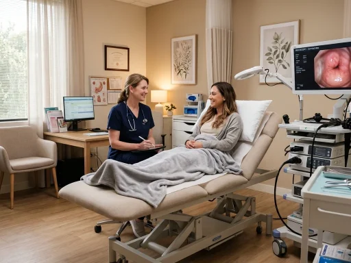
- Diagnostic Office Hysteroscopy: The direct visual inspection of the endocervical canal, internal os, and endometrium offers unmatched diagnostic precision, enabling the surgeon to establish an accurate surgical plan before entering the operating suite.
Get our latest guides, news, and insights highlighted in your Google Search & AI Overviews.