
✨ This article was AI edited. Editorial responsibility: Hysteroscopy.info.
Produced with AI. This article was artificially generated. Editorial responsibility: Hysteroscopy.info.
A diagnostic hysteroscopy typically takes 5 to 15 minutes, while an operative hysteroscopy (such as polyp or fibroid removal) generally lasts 15 to 45 minutes. Total clinic time ranges from 1 to 3 hours including preparation, cervical anesthesia, the endoscopic procedure, and post-sedation recovery observation.
Understanding the exact time commitment for a hysteroscopy helps alleviate procedural anxiety and allows for proper scheduling of work leave, transportation, and post-operative rest. The duration of the procedure depends significantly on whether the intervention is purely diagnostic or operative, whether cervical dilation is required, the type of anesthesia administered, and the clinical setting (outpatient clinic vs. hospital surgical suite).
Diagnostic vs. Operative Hysteroscopy: Duration Comparison
Hysteroscopy is divided into two primary procedural classifications, each carrying distinct temporal parameters and technical requirements:
| Procedure Type | Intrauterine Scope Time | Total Operating Room Time | Total Clinic / Facility Stay |
|---|---|---|---|
| Diagnostic Hysteroscopy (In-Office) | 5 – 10 minutes | 15 – 25 minutes | 45 – 90 minutes |
| Diagnostic with Endometrial Biopsy | 8 – 15 minutes | 20 – 30 minutes | 60 – 90 minutes |
| Operative Hysteroscopy: Polypectomy | 15 – 30 minutes | 35 – 50 minutes | 2 – 3 hours |
| Operative Hysteroscopy: Myomectomy (Fibroid Resection) | 25 – 45 minutes | 45 – 75 minutes | 3 – 4 hours |
| Hysteroscopic Lysis of Adhesions (Asherman’s) | 20 – 40 minutes | 40 – 60 minutes | 2.5 – 3.5 hours |
Phase-by-Phase Procedural Timeline
To accurately plan your day, consider the chronological progression of an outpatient hysteroscopy appointment from arrival to discharge:
Phase 1: Pre-Procedure Intake & Preparation (30 – 45 Minutes)
Upon arriving at the clinical facility, the pre-operative sequence begins:
- Patient Registration & Vitals: Confirmation of identity, medical history verification, and baseline vital signs recording (blood pressure, heart rate, oxygen saturation).
- Pre-Operative Medication: Administration of oral nonsteroidal anti-inflammatory drugs (NSAIDs such as Ibuprofen 800mg) or anxiolytics if prescribed to mitigate uterine cramping.
- Cervical Priming Assessment: If cervical ripening agents (such as vaginal Misoprostol) were prescribed the previous night, the clinician verifies adequate cervical softening.
- Bladder Evacuation: Emptying the bladder to optimize pelvic anatomy orientation and comfort during speculum insertion.
Phase 2: Positioning & Cervical Anesthesia (5 – 10 Minutes)
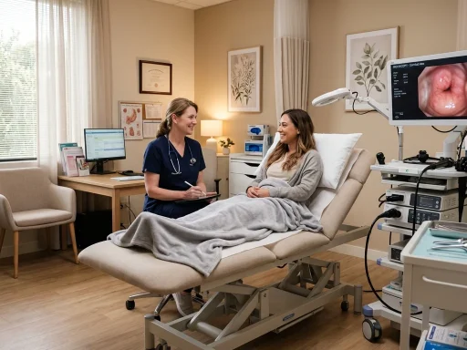
Once escorted to the examination room, positioning and local anesthesia administration proceed as follows:
- Lithotomy Positioning: The patient is positioned comfortably in standard dorsal lithotomy position on the gynecological table.
- Speculum Placement & Antiseptic Cleansing: A bivalve speculum is gently placed, and the cervix is cleansed with an antiseptic povidone-iodine or chlorhexidine solution.
- Paracervical or Intracervical Block: For patients receiving local anesthesia, 1% to 2% lidocaine is injected around the cervix. The onset of local nerve block takes approximately 2 to 3 minutes.
Phase 3: The Endoscopic Procedure (5 – 30 Minutes)
The active endoscopic phase consists of gentle instrument insertion, continuous fluid distension, and cavity inspection:
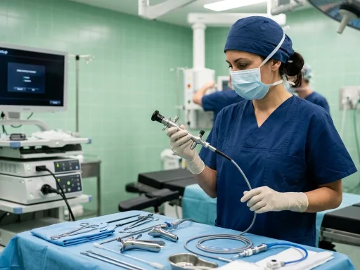
- Hysteroscope Insertion: A miniature, illuminated telescope (ranging from 2.9mm to 5.0mm in outer diameter) is guided through the cervical canal into the uterine cavity without the need for aggressive mechanical dilation in most diagnostic cases.
- Fluid Distension: Sterile normal saline solution is continuously infused through the inflow sheath under controlled intrauterine pressure (typically 70 to 100 mmHg). This gently separates the anterior and posterior endometrial walls for clear optical visualization.
- Cavity Survey & Targeted Interventions: The gynecologist systematically inspects the endocervical canal, uterine fundus, anterior/posterior walls, and right and left tubal ostia. If pathological lesions (such as endometrial polyps or submucosal myomas) are identified, micro-instruments, bipolar electrodes, or mechanical tissue extraction devices (e.g., MyoSure) are introduced through the operating channel to resect the tissue.
Phase 4: Post-Procedure Observation & Discharge (15 – 60 Minutes)
After the hysteroscope is withdrawn, post-operative monitoring commences:
- In-Office Procedures without Sedation: Patients typically rest in the procedure room for 10 to 15 minutes. Once vital signs are stable and cramping has subsided, patients may dress and walk out independently.
- Hospital or Ambulatory Center with Sedation (Monitored Anesthesia Care / General): Patients are transferred to the Phase I Post-Anesthesia Care Unit (PACU) for 45 to 60 minutes until alert, tolerating fluids, and capable of ambulation. An accompanying adult is legally required to drive the patient home.
Factors That Influence Procedure Duration
Several clinical and anatomical factors can extend or shorten the surgical time:
- Cervical Stenosis: A tight or scarred external or internal cervical os may require incremental mechanical dilation using Hegar or Pratt dilators, adding 5 to 10 minutes to the procedure.
- Pathology Size and Number: Resecting a single 1cm pedunculated endometrial polyp takes roughly 3 to 5 minutes, whereas morcellating a 3cm broad-based Type II submucosal fibroid may require 30 to 45 minutes of meticulous surgical technique.
- Distension Fluid Deficit Limits: Strict fluid balance monitoring is mandatory during operative hysteroscopy. If isotonic saline absorption reaches predetermined safety thresholds (e.g., 2,500 mL in healthy premenopausal patients), the procedure must be paused or concluded to prevent fluid overload syndromes.
- Uterine Flexion & Anatomical Anomalies: Severe uterine retroflexion or congenital uterine septa require cautious navigation to avoid uterine wall perforation.
Expected Recovery Milestones by Hour and Day
Post-procedure recovery is remarkably rapid compared to traditional abdominal or laparoscopic surgeries:
- Hour 0 – 2: Mild to moderate uterine cramping (similar to menstrual cramps) and light watery or blood-tinged discharge. Manageable with oral NSAIDs and a heating pad.
- Day 1 (24 Hours): Most patients who had in-office diagnostic hysteroscopy return to desk work, light household tasks, and routine activities the following morning.
- Days 2 – 7: Residual light spotting or pinkish discharge is normal as the endometrium regenerates. Pelvic rest (avoiding tampons, intercourse, and swimming) is recommended for 1 to 2 weeks.
Frequently Asked Questions
Can I drive myself home after a hysteroscopy?
If your hysteroscopy is performed in-office with local anesthesia only, you are generally safe to drive yourself home after a short observation period. However, if any oral sedative, nitrous oxide, IV conscious sedation, or general anesthesia is administered, you must arrange for a responsible adult to escort and drive you home.
How much time should I take off from work?
For a diagnostic hysteroscopy, taking the day of the procedure off is standard, with most individuals returning to work the next morning. For operative hysteroscopy involving fibroid removal or extensive polyp resection under sedation, taking 24 to 48 hours off is recommended to allow full recovery from fatigue and mild cramping.
Why does an operative hysteroscopy take longer than a diagnostic one?
Operative hysteroscopy requires the precise assembly and introduction of surgical energy or mechanical tissue extraction tools, continuous fluid balance measurement, meticulous tissue resection, and verification of complete hemostasis before scope removal.
For a full post-procedure recovery protocol, explore our comprehensive Week-by-Week Hysteroscopy Recovery Guide and our detailed Step-by-Step Hysteroscopy Experience Guide.
Get our latest guides, news, and insights highlighted in your Google Search & AI Overviews.