
✨ This article was AI edited. Editorial responsibility: Hysteroscopy.info.
Produced with AI. This article was artificially generated. Editorial responsibility: Hysteroscopy.info.
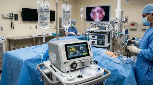
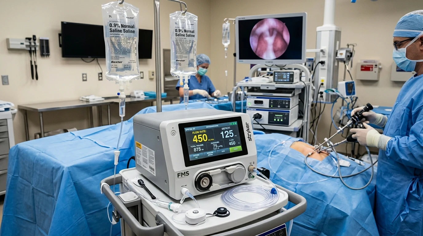
Hysteroscopy fluid is sterile distension liquid—primarily isotonic 0.9% normal saline—infused into the uterus to expand the cavity and clear blood for optical visualization. Automated fluid management systems continuously monitor intrauterine pressure (70–100 mmHg) and calculate fluid absorption deficit, capping absorption at 2,500 mL for saline to strictly prevent fluid overload complications.
Because the uterine cavity is normally a collapsed potential space with opposing muscular walls, optical endoscopy requires continuous fluid distension to separate the walls and wash away debris. Intraoperative fluid management is the cornerstone of patient safety during diagnostic and operative hysteroscopy, governed by strict thermodynamic and fluid balance principles.
Types of Hysteroscopic Distension Media
Distension media are divided into two main categories based on electrical conductivity and osmolarity:
| Distension Media | Osmolarity & Composition | Electrosurgical Compatibility | Maximum Safe Fluid Deficit | Clinical Application & Safety Notes |
|---|---|---|---|---|
| 0.9% Normal Saline (NaCl) (Isotonic Electrolyte) | Isotonic (308 mOsm/L); contains sodium & chloride | Compatible with Bipolar Electrosurgery and Mechanical Morcellators | 2,500 mL (in healthy premenopausal patients) | The international gold standard; eliminates the risk of acute hyponatremia and cerebral edema. |
| Lactated Ringer’s Solution (Isotonic Electrolyte) | Isotonic (273 mOsm/L); balanced electrolyte solution | Compatible with Bipolar Electrosurgery | 2,500 mL | Safe physiological electrolyte profile; alternative to normal saline. |
| 1.5% Glycine (Hypotonic Non-Electrolyte) | Hypotonic (200 mOsm/L); amino acid derivative | Compatible with Monopolar Electrosurgery only | 1,000 mL (Strict threshold) | Historical medium; excessive absorption causes acute hyponatremia, volume overload, and hyperammonemia. |
| 3% Sorbitol / 5% Mannitol (Hypotonic Non-Electrolyte) | Hypotonic / Isosmolar non-electrolytic sugar alcohols | Compatible with Monopolar Electrosurgery only | 1,000 mL | Non-conductive sugar solutions; carries risk of osmotic diuresis and hyponatremia upon vascular intravasation. |
Hydrostatic Pressure Dynamics and Intravasation Physiology
Fluid distension operates under a fundamental hemodynamic threshold:
- Mean Arterial / Venous Pressure Threshold: Intravasation (the systemic absorption of fluid into the patient’s bloodstream) begins when intrauterine distension pressure exceeds the patient’s Mean Uterine Venous Pressure, typically between 70 mmHg and 80 mmHg.
- Target Operating Pressure: Gynecologists maintain distension pressure between 70 mmHg and 100 mmHg (or just below the mean arterial pressure), providing clear optical views while minimizing rapid venous uptake through opened myometrial sinuses.
Automated Fluid Management Systems (FMS)
Modern surgical suites utilize automated electronic fluid management systems (such as the Stryker, Olympus, or Hologic systems) equipped with precision load-cell scales. These machines continuously calculate:
If the fluid deficit approaches critical warning thresholds (e.g., 1,500 mL for saline or 750 mL for hypotonic media), auditory alarms alert the surgical team to expedite resection, lower infusion pressure, or conclude the procedure.
Clinical Protocol for Managing High Fluid Deficit
When fluid deficit reaches predetermined clinical thresholds:
- At 1,500 mL Saline Deficit: The anesthesiologist is notified, intrauterine pressure is decreased, and a completion timeline is established.
- At 2,500 mL Saline Deficit (or 1,000 mL Non-Electrolyte Deficit): The procedure is immediately halted, distension fluid is evacuated, and IV diuretics (e.g., Furosemide) and serum electrolyte panels are ordered if clinically indicated.
Frequently Asked Questions
Why does watery fluid leak out after my hysteroscopy?
It is completely normal to experience watery, pink-tinged fluid drainage for 24 to 48 hours following a hysteroscopy. This represents residual sterile saline draining from the uterine cavity and vaginal vault as tissues settle.
How does modern bipolar hysteroscopy prevent fluid overload dangers?
Bipolar instruments utilize normal saline containing natural body-compatible sodium concentrations. Unlike older monopolar systems requiring glycine, systemic absorption of saline does not dilute blood sodium levels, virtually eliminating the danger of hyponatremic encephalopathy (TUR syndrome).
How is fluid safety monitored during my surgery?
A dedicated circulating nurse and automated computerized scales monitor fluid intake and collection in real time every few minutes throughout the operation.
Learn more about procedural safety in our Step-by-Step Procedure Guide and explore our Day-by-Day Recovery Timeline.
Thermodynamic Considerations: Fluid Temperature and Patient Comfort
In addition to volume and pressure regulation, the thermodynamic properties of distension media play a crucial clinical role during hysteroscopy:
- Warming Distension Saline: Warming normal saline to normal physiological body temperature (37°C / 98.6°F) prior to infusion significantly reduces intraoperative uterine vasospasm, shivering, and patient-reported cramping pain compared to room-temperature fluids.
- Prevention of Intraoperative Hypothermia: In extended operative procedures where larger volumes of fluid are circulated, utilizing automated fluid warming sleeves prevents systemic core body temperature drop during general anesthesia.
Clinical Guidelines from International Gynecological Societies
Both the American College of Obstetricians and Gynecologists (ACOG) and the European Society for Gynaecological Endoscopy (ESGE) maintain strict consensus guidelines for intrauterine fluid safety:
- Routine Intraoperative Documentation: The circulating nursing staff must verbally announce and document fluid deficit totals at minimum 10-minute intervals during operative resection.
- Pre-Operative Renal & Cardiac Screening: Patients undergoing extensive myomectomy or resection of deep intrauterine lesions must be screened for underlying cardiopulmonary or renal impairments that could alter fluid tolerance.
- Emergency Response Preparedness: Surgical teams must have immediate access to IV diuretics (Furosemide 20–40mg) and hypertonic saline (3% NaCl) in the rare event of severe non-electrolyte fluid intravasation.
Comparative Summary: Choosing the Right Distension System
Modern clinical hysteroscopy prioritizes isotonic normal saline paired with advanced automated pump systems for virtually all diagnostic and operative indications. By eliminating the risks of severe electrolytic imbalances associated with historical monopolar distension media, clinicians can safely perform complex intrauterine procedures with maximal peace of mind and rapid patient recovery.
Get our latest guides, news, and insights highlighted in your Google Search & AI Overviews.