
✨ This article was AI edited. Editorial responsibility: Hysteroscopy.info.
Produced with AI. This article was artificially generated. Editorial responsibility: Hysteroscopy.info.
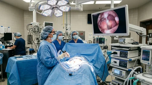
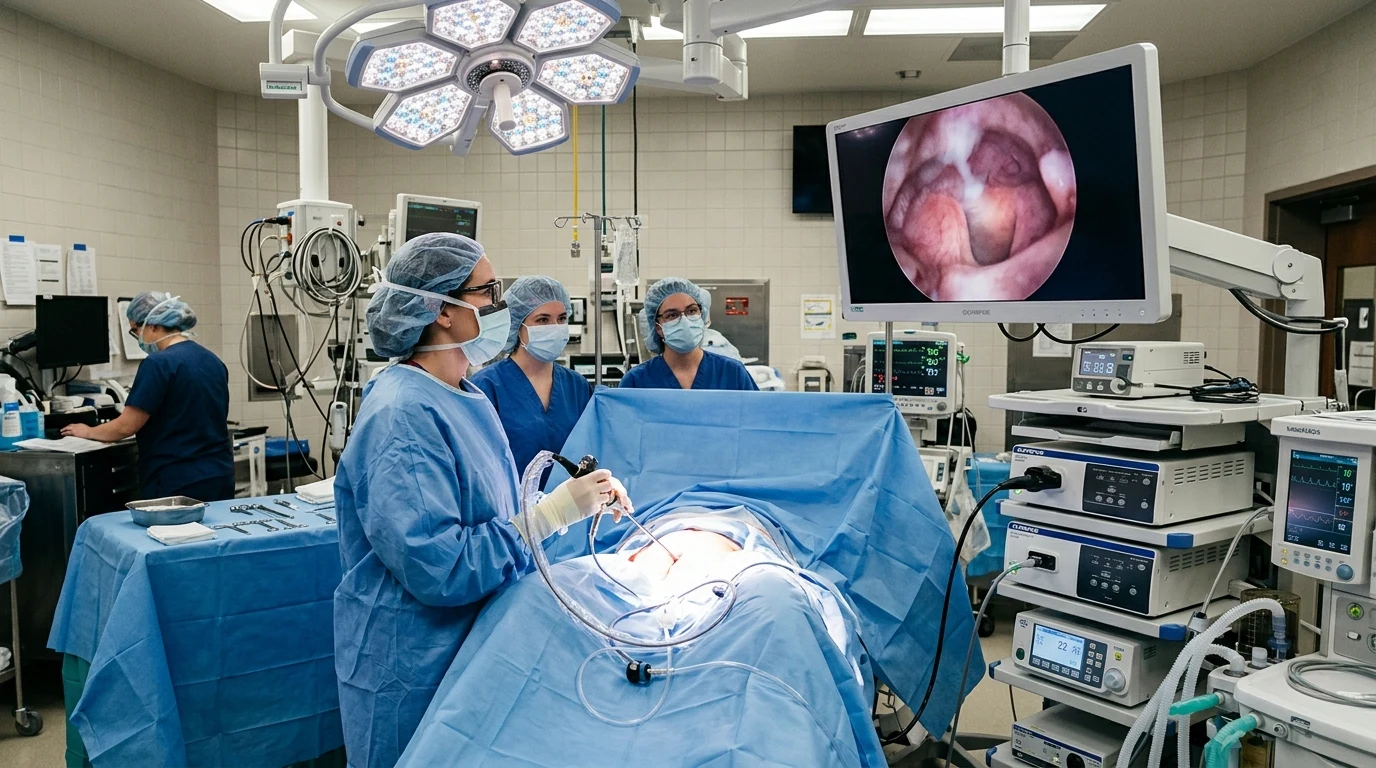
Hysteroscopic myomectomy is a transcervical, incision-free surgical procedure designed to resect submucosal uterine fibroids protruding into the endometrial cavity. Utilizing a bipolar resectoscopic loop or mechanical tissue extraction system, surgeons slice and extract fibroid tissue under continuous fluid distension in 20 to 45 minutes, resolving menorrhagia and enhancing fertility.
Submucosal uterine leiomyomas represent the most clinically disruptive variant of uterine fibroids. Despite accounting for only a fraction of total fibroid volume, their direct contact with and distortion of the endometrial lining causes profound menorrhagia, iron-deficiency anemia, dysmenorrhea, and reproductive failure. Hysteroscopic myomectomy provides the gold standard uterus-preserving solution without requiring abdominal incisions or prolonged hospital stays.
FIGO Classification and Surgical Selection
Procedural planning is dictated by the International Federation of Gynecology and Obstetrics (FIGO) classification of submucosal fibroids:
| FIGO Subtype | Intramural Extension | Intracavitary Protrusion | Surgical Feasibility & Approach |
|---|---|---|---|
| Type 0 | 0% (Pedunculated) | 100% within the cavity | Single-stage hysteroscopic resection; highly straightforward excision. |
| Type 1 | < 50% intramural extension | ≥ 50% intracavitary protrusion | Standard hysteroscopic myomectomy; excellent single-stage completion (>90%). |
| Type 2 | ≥ 50% intramural extension | < 50% intracavitary protrusion | Advanced hysteroscopic surgery; often requires planned 2-stage resection. |
Surgical Modalities: Loop Resectoscopy vs. Mechanical Morcellation
Gynecological surgeons employ two modern technological platforms for intrauterine fibroid removal:
1. Classical Bipolar Loop Resectoscopy
Utilizes a motorized semicircular wire loop operating with high-frequency radiofrequency energy within isotonic normal saline. The surgeon systematically shaves the fibroid tissue from the apex down to the pseudocapsule in progressive thin strips, providing immediate coagulation of feeding myometrial vessels.
2. Mechanical Tissue Removal Systems (e.g., MyoSure, TruClear)
An innovative mechanical approach employing a high-speed rotating and reciprocating blade housed within a slender cannula. As tissue is shaved mechanically, continuous vacuum suction pulls the fragments through the scope directly into an external specimen container. This eliminates repeated scope removals, reduces operative duration, and avoids thermal injury to the surrounding endometrial lining.
Fluid Deficit Monitoring and Patient Safety
Continuous infusion of normal saline creates an optical surgical field. During myometrial resection, venous sinuses absorb distension fluid into the systemic circulation. Intraoperative safety protocols enforce:
- Automated Fluid Balance Scales: Computerized systems measure fluid inflow and outflow continuously.
- Deficit Threshold Caps: For isotonic saline in healthy premenopausal patients, fluid absorption is strictly capped at 2,500 mL. Reaching this threshold requires immediate cessation of the procedure to prevent pulmonary edema or fluid overload.
Post-Operative Recovery and Fertility Outcomes
Because no cutaneous incisions are made, recovery is remarkably rapid:
- First 24 Hours: Mild lower pelvic cramping and light reddish discharge. Discharged home within 1 to 2 hours with oral NSAIDs.
- Days 2 – 5: Return to desk work, light walking, and daily routines. Avoid heavy lifting (>15 lbs).
- Weeks 1 – 2: Resolution of cramping; tapering pinkish discharge as the myometrial bed re-epithelializes. Pelvic rest observed for 2 weeks.
- Fertility Prognosis: Resection of Type 0 and Type 1 submucosal fibroids increases subsequent clinical pregnancy rates by 40% to 50% in women experiencing subfertility.
Frequently Asked Questions
How long does a hysteroscopic myomectomy take?
The active surgical resection takes approximately 20 to 45 minutes, with total facility stay lasting 2 to 4 hours including pre-op preparation and post-anesthesia recovery.
Will hysteroscopic myomectomy weaken the uterine wall for future pregnancy?
Because the outer myometrium and uterine serosa are not cut, hysteroscopic myomectomy maintains excellent uterine integrity, and patients are generally eligible for normal vaginal deliveries in future pregnancies.
Can fibroids grow back after being removed?
The specific fibroid tissue resected is permanently eliminated. However, new fibroids can potentially form in other areas of the myometrium over time, with a 5-year recurrence rate of 10% to 15%.
For more recovery details, explore our Complete Day-by-Day Recovery Timeline and read our guide on CPT Coding and Insurance Guide.
Intraoperative Complication Prevention and Safety Pearls
Ensuring absolute surgical safety during hysteroscopic fibroid resection requires strict adherence to international surgical guidelines:
- Uterine Wall Thickness Assessment: The surgeon maintains at least 5mm of intact myometrial mantle between the resection base and the outer serosa, monitored via real-time ultrasound if the intramural component is deep.
- Minimizing Operating Time: Operative duration is kept under 60 minutes to reduce fluid intravasation and surgical fatigue.
- Electrosurgical Grounding Protocols: True bipolar energy circuits confine current flow exclusively between the two active poles at the loop tip, eliminating stray current thermal burns to adjacent pelvic organs.
- Cervical Priming Agents: Pre-operative administration of vaginal misoprostol or osmotic dilators softens the cervix, preventing mechanical lacerations during resectoscope insertion.
Long-Term Quality-of-Life and Bleeding Relief
Clinical data confirms that over 90% of women achieve complete resolution of menorrhagia and iron-deficiency anemia within 3 months following hysteroscopic myomectomy, restoring full physical energy and vitality without need for chronic hormonal suppressive medications.
Anesthesia Modalities and Intraoperative Monitoring
Anesthesia plans are customized based on the FIGO fibroid classification and patient health profile:
- Monitored Anesthesia Care (MAC) with Paracervical Block: Ideal for Type 0 pedunculated polyps and fibroids, enabling rapid cognitive recovery and same-day discharge within 60 minutes.
- General Laryngeal Mask Anesthesia: Recommended for complex Type 2 submucosal myomas to ensure absolute immobility during delicate micro-loop resection.
Key Clinical Takeaways for Patients
Modern hysteroscopic procedures represent the pinnacle of conservative gynecological care, combining high-resolution optical precision with gentle tissue preservation. Following your individualized post-operative instructions, taking recommended anti-inflammatory medications, and attending your scheduled follow-up visits ensures rapid recovery and optimal reproductive health.
Always communicate openly with your surgical team regarding any post-operative questions or unusual symptoms.
Get our latest guides, news, and insights highlighted in your Google Search & AI Overviews.