
✨ This article was AI edited. Editorial responsibility: Hysteroscopy.info.
Produced with AI. This article was artificially generated. Editorial responsibility: Hysteroscopy.info.
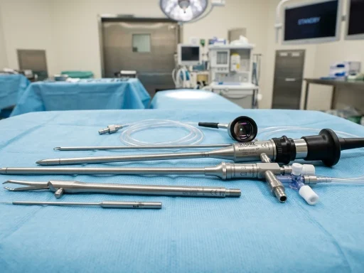
A hysteroscopy to remove fibroids (hysteroscopic myomectomy) is an outpatient surgical procedure performed through the vagina and cervix to excise submucosal fibroids without abdominal incisions. Using a specialized resectoscope or tissue extraction morcellator, the surgeon shaves and removes intracavitary fibroids in 20 to 45 minutes under general or local anesthesia.
Uterine fibroids (leiomyomas) that grow directly beneath the endometrial lining and protrude into the uterine cavity are classified as submucosal fibroids. These lesions are a leading cause of severe menorrhagia (abnormally heavy, prolonged menstrual bleeding), debilitating cramps, chronic iron-deficiency anemia, and reproductive complications. Performing a hysteroscopy to remove fibroids provides immediate therapeutic relief, restores uterine anatomical integrity, and preserves reproductive capability.
Who Is a Candidate for Hysteroscopic Fibroid Removal?
Candidate eligibility is determined by the size, depth, and cavity protrusion of the fibroid as characterized by transvaginal ultrasound, saline infusion sonohysterography (SIS), or pelvic MRI:
| Submucosal Category | Anatomical Location | Ideal Fibroid Diameter | Procedural Success Rate |
|---|---|---|---|
| FIGO Type 0 (Pedunculated) | 100% inside uterine cavity | 0.5 cm – 5.0 cm | >95% complete single-session removal |
| FIGO Type 1 (Sessile) | >50% intracavitary / <50% intramural | 1.0 cm – 4.0 cm | 90% – 95% single-stage excision |
| FIGO Type 2 (Deep Submucosal) | <50% intracavitary / >50% intramural | 1.0 cm – 3.5 cm | 80% – 85% (may require staged 2-step procedure) |
Step-by-Step Surgical Technique
The surgical protocol for hysteroscopic fibroid removal follows a precise, standardized sequence designed to maximize tissue clearance while protecting the surrounding endometrial lining:
1. Anesthesia and Cervical Dilation
The patient receives intravenous general anesthesia or deep conscious sedation paired with a local paracervical block. The cervix is gently dilated using graduated Hegar dilators up to 9mm to 10mm to accommodate the outer sheath of the operating resectoscope.
2. Uterine Distension & Cavity Assessment
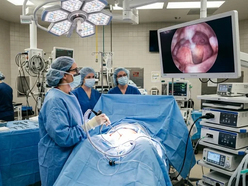
The resectoscope is introduced under direct optical visualization while sterile normal saline is infused under regulated hydrostatic pressure. The surgeon performs a panoramic inspection of the cavity to confirm the fibroid’s base diameter, vascularity, and relationship to the tubal ostia.
3. Progressive Layered Resection (Loop Resectoscopy or Mechanical Morcellation)
Using a bipolar wire loop electrode, the surgeon slices the fibroid systematically from top to base in thin chips, achieving simultaneous hemostasis. Alternatively, a high-speed mechanical tissue removal device (such as MyoSure) is placed directly against the fibroid, simultaneously shaving and suctioning the tissue fragments out of the uterus.
4. Safety Fluid Balance Verification & Scope Withdrawal
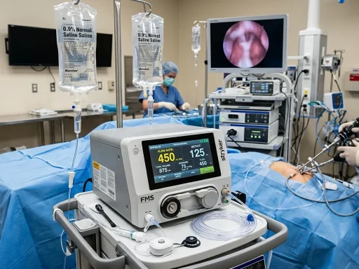
Throughout the procedure, automated fluid management scales monitor fluid deficit. Once the fibroid is completely resected flush with the surrounding myometrial pseudocapsule and hemostasis is confirmed, the distension medium is drained, and the instrument is gently withdrawn.
Post-Operative Recovery and What to Expect
Because hysteroscopic fibroid resection requires no abdominal cuts, recovery milestones are remarkably swift compared to open or laparoscopic myomectomies:
- First 24 Hours: Mild lower pelvic cramping and light reddish-brown discharge. Manageable with oral ibuprofen and acetaminophen. Most patients are discharged home within 1 to 2 hours of surgery.
- Days 2 – 5: Transition to normal daily activities, light walking, and desk work. Avoid strenuous cardiovascular exercise and heavy lifting (>15 lbs).
- Weeks 1 – 3: Tapering watery or pinkish discharge as the myometrial resection bed heals and re-epithelializes. Strict pelvic rest (no tampons, no intercourse, no tub baths) is observed for 2 weeks.
- First Menstrual Period: Typically occurs 4 to 6 weeks post-procedure and is notably lighter and less painful than pre-operative periods.
Clinical Benefits and Symptom Relief
Removing submucosal fibroids hysteroscopically achieves transformative improvements across key quality-of-life and clinical metrics:
- Bleeding Control: Over 90% of women report a dramatic reduction in menstrual flow, eliminating heavy clots and flooding.
- Anemia Resolution: Hemoglobin and serum ferritin levels rapidly normalize within 2 to 3 months without ongoing need for blood transfusions or iron infusions.
- Fertility Enhancement: Eliminating the intracavitary mass effect increases spontaneous conception rates and significantly enhances IVF embryo implantation success rates by 40% to 50%.
Frequently Asked Questions
How long does a hysteroscopy to remove fibroids take?
The active surgical resection typically takes 20 to 45 minutes, with total operating room and recovery time lasting approximately 2 to 3 hours.
Will removing fibroids hysteroscopically leave scars on my abdomen?
No. Hysteroscopic fibroid removal is performed entirely transcervically through the natural opening of the cervix. There are zero skin incisions and zero external scars.
Can all uterine fibroids be removed with hysteroscopy?
No. Hysteroscopy is specifically designed for submucosal fibroids that project into the uterine cavity (FIGO Types 0, 1, and 2). Intramural fibroids (located entirely within the muscle wall) or subserosal fibroids (growing on the outside of the uterus) require laparoscopic, robotic, or abdominal surgical approaches.
For more detailed recovery timelines, explore our Complete Day-by-Day Recovery Timeline and read our overview on Hysteroscopy vs. Laparoscopy Options.
Comparative Analysis: Resectoscopic Loop vs. Mechanical Morcellation
When selecting the optimal surgical approach for submucosal fibroid excision, gynecologists weigh the distinct advantages of resectoscopic loop electrocautery versus modern mechanical tissue extraction systems:
| Feature / Metric | Classical Bipolar Resectoscope | Mechanical Tissue Removal (e.g., MyoSure) |
|---|---|---|
| Energy Source | Bipolar High-Frequency Radiofrequency | Non-thermal Mechanical Blade with Suction |
| Operating Time (Average 2cm Fibroid) | 30 – 45 minutes | 10 – 20 minutes |
| Thermal Spread Risk | Minimal, but present at myometrial base | Zero thermal spread to healthy endometrium |
| Specimen Collection Method | Manual chip retrieval via forceps/curette | Continuous automated vacuum canister collection |
| Ideal Clinical Scenario | Fibroids with deep vascular pedicles needing coagulation | Submucosal polyps and soft-to-firm Type 0/1 leiomyomas |
Long-Term Reproductive and Quality-of-Life Outcomes
Longitudinal clinical trials evaluating patients 12 to 36 months following hysteroscopic fibroid resection document enduring symptom resolution and high patient satisfaction rates (>92%). Over 85% of women achieve sustained normalization of menstrual bleeding patterns, reducing chronic fatigue and restoring vitality.
Anesthesia Options and Pain Management Protocols
Tailoring the anesthetic plan ensures maximal safety and rapid outpatient discharge:
- General Endotracheal Anesthesia: Preferred for complex Type 2 submucosal fibroids or cases requiring concurrent laparoscopic assessment.
- Intravenous Monitored Anesthesia Care (MAC) with Local Block: Provides deep sedation and rapid cognitive recovery for straightforward Type 0 and Type 1 myomectomies.
- Post-Operative Analgesia: Multimodal non-opioid regimens combining scheduled oral ibuprofen (600mg every 6 hours) with acetaminophen (1000mg every 8 hours) provide superior cramping control without sedation or nausea.
Get our latest guides, news, and insights highlighted in your Google Search & AI Overviews.